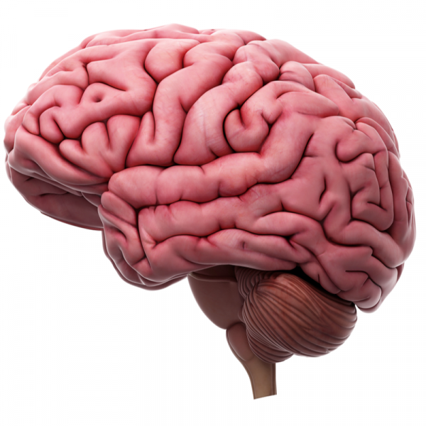
What are arachnoid cysts?
An arachnoid cyst is located in the arachnoid, the spider tissue and innermost covering layer of the brain. It is a benign cavity that is filled with neural fluid. An arachnoid cyst can develop anywhere in the brain and is usually congenital. Almost 50 per cent of all arachnoid cysts occur in the area of the Sylvian fissure (extended lateral furrow of the brain). 10 per cent of all arachnoid cysts are located suprasellar (in the third cerebral ventricle), in the pineal recess (in the area of the epithalamu) or above the cerebellar vermis. 5 per cent of all arachnoid cysts are interhemispheric (within a cerebellar or cerebral hemisphere). Less frequently, arachnoid cysts occur in other brain regions.
What are the symptoms of an arachnoid cyst?
The arachnoid cyst is only associated with symptoms in about 10 to 20 percent of all cases, which can be explained by the pressure of the cyst on the surrounding brain tissue. Headaches are among the most common complaints. More rarely, hormonal disorders or seizures can also be caused by an arachnoid cyst. However, reduced vision and/or uncontrolled head wobbling, which occurs from time to time, can also be signs of an arachnoid cyst and should definitely be clarified by a doctor.
How is an arachnoid cyst diagnosed?
An arachnoid cyst is often discovered by chance during an examination of the head. The usual imaging methods are computer tomography (CT) or magnetic resonance imaging (MRI). If the suspicion of an archnoid cyst is confirmed, an MRI can show the cyst in three planes and thus clearly show its location in relation to important anatomical structures such as the optic nerve, the aorta and the brain chambers (ventricles). This precise knowledge of the localisation of the archnoid cyst is of great importance for therapy planning.
How is an archnoid cyst treated?
If it is an archnoid cyst that causes symptoms, surgical removal can be considered. In most cases, asymptomatic archnoid cysts do not even require therapy. However, the archnoid cyst should be checked regularly by a doctor using the usual imaging procedures.
If a relatively large archnoid cyst is discovered in a child, surgical intervention is usually recommended so that the brain has enough space for normal development. There are various surgical procedures available to the doctor for this. As a rule, the endoscopic method attempts to drain the cyst contents into the basal cisterns or into the brain chambers (ventricles). If the endoscopic method is not promising for anatomical reasons, microsurgery can also be used.
| Pathogen | Source | Members - Area |
|---|---|---|
| Arachnoid cysts | EDTFL | As a NLS member you have direct access to these frequency lists |
| Arachnoid diverticulum | EDTFL | As an NLS member you have direct access to these frequency lists |
