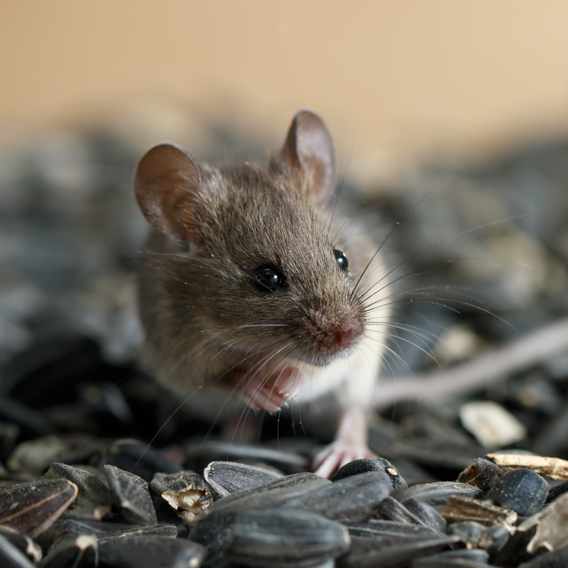
What is the Bartonella taylorii pathogen?
Bartonella taylorii belongs to the Bartonella genus and is a Bacterium. Like other Bartonella species, the Bartonella taylorii pathogen can cause disease in animals. A study of small mammals from Turkish Thrace has revealed that rodents in particular play a crucial role as reservoir for B. taylorii. The prevalence of Bartonella taylorii pathogens is particularly pronounced in the swamp forest habitat.
How was the Bartonella taylorii pathogen diagnosed in rodents?
The Bartonella taylorii pathogen was first detected in rodents in the Turkish Trakien. For this purpose, ninety rodents, belonging to three different rodent species, were screened by PCR . 22.2 percent of the rodents were the total prevalence of Bartonella infections. Based on the phylogenetic analyses of the two household genes rpoB and gltA, the strains were molecularly characterised.
What symptoms did the rodents exhibit due to Bartonella taylorii infection?
One raccoon showed clinical signs of tremors and weakness. After the animal was euthanised, the autopsy revealed a generally poor body condition. This poor general condition manifested itself in the animal as follows:
- a diffuse palpable enlargement of the lymph node (lymphadenopathy),
- pale, firm kidneys, which were interspersed with petechial haemorrhages throughout the renal cortex,
- histological lesions such as systemic fibrinoid circulatory disturbances of the vessels (vascular necrosis),
- severe renal lesions with lymphoplasmacytic interstitial nephritis (inflammation of the renal tubules),
- fibrinosuppurative glomerulonephritis.
In the raccoon, inflammatory vascular
lesions were also found within the uvea, the heart as well as the lymph nodes and the
lamina propria of the stomach wall. Additional diagnostic tests,
performed, were negative for the following pathogens:
- the Bacterium Borrelia burgdorferi,
- the Bacterium Leptospira,
- Aleutian disease virus,
- Canine distemper virus, also called canine distemper virus,
- feline coronavirus, which primarily affects cats,
- porcine circovirus 2 (DNA virus from the Circoviridae family),
- Rabies virus.
What are the histological characteristics of the Bartonella taylorii pathogen?
Transmission electron microscopy revealed a large group of bacterial rods approximately 1.3 × 0.35 µm in size. These showed a trilaminar cell wall located within the glomeruli. Analysis of partial 16S-23S ribosomal RNA intergenic spacer region sequences from kidney tissue and the citrate synthase gene confirmed that the B. taylorri pathogen is related to numerous Bartonella species.
In which rodents could the Bartonella taylorii pathogen be detected?
The Bartonella taylorii pathogen has been detected in a variety of Eurasian rodent and flea species. The pathogen has also been detected in shrews in Europe . In North America, the Bartonella taylorii pathogen could be diagnosed in raccoons.
What infection does the Bartonella taylorii pathogen cause in mice?
In a study, it was investigated which high-grade infection the Bartonella taylorii pathogen can cause in immunocompromised mice. For this purpose, the animals were infected with the Bacterium and then observed. Superficial observations revealed that two months after infection, no abnormalities could be detected in the animals. After four months, however, all mice showed clearly enlarged spleens.
A more detailed examination under a light microscope revealed several pathologies in various organs at an earlier stage. For example, about one month after infection, a sparse myeloid infiltrate was diagnosed in the liver. This myeloid infiltrate was mainly neutrophils as well as band forms, which also appeared as nests of cells. Researchers believe that these inflammatory cells were either microabscesses (neutrophils only) or foci of extramedullary haematopoiesis. The inflammatory cells became more prominent again at a later time.
Furthermore, a neutrophilic and mononuclear infiltrate around the central nerves was observed between the first and second month after infection. This could indicate mild hepatitis. After three months, spindle-shaped cells and foci were detected. These lesions appeared to invade the surrounding liver tissue with no clear boundaries. If the lesions were smaller, they often had a border of neutrophils (subtype of white blood cells) and mononuclear (cells with a single nucleus) cells. The sinusoids adjacent to the lesions were often dilated. After four months of infection, the lesions accounted for about half of the normal liver tissue and some also had calcifications. Remnants of maturing myeloid cells and megakaryocytes were also present in the entire non-lesional area of the liver tissue. Around the same time, lesions with a similar external appearance could also be detected in the kidney. This indicated the form of granulomatous nephritis.
Within the study, it could thus be demonstrated that the Bartonella taylorii pathogen can cause a series of chronic, high-grade infections in immunocompromised mice. The pathologies, caused by the pathogen, resembled the features that can also be observed in immunocompromised patients after they have developed a Bartonella infection.
Why is further research into the Bartonella taylorii pathogen urgently needed?
Rodents are ubiquitous in urban environments. Through easy contact with humans, it is imperative that veterinarians, as well as medical professionals, continue research on the pathogen. The pathogen should also be given more attention in medical examinations as it is usually difficult to detect through a routine bacterial culture. In rodents with the symptoms described above, additional molecular tests should therefore be performed to detect the pathogen.
