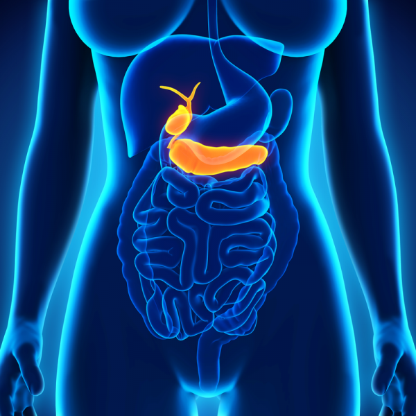
What is a Choldedochal cyst?
A Choldedochal cyst describes the cystic dilatation of the (extrahepatic) bile ducts located outside the liver and can be summarised briefly under the phenomenon of bile duct dilatation.
Cyst: Greek kystis "bladder"=a tissue cavity closed off by an epithelium (membrane)
What can cause cholledochal cysts?
Doctors are still unclear about the exact causes of a cholledochal cyst (bile duct cyst). However, they assume that the excretory duct of the pancreas forms a common channel with the bile ducts, which deviates from the norm in this way. This common channel ensures that pancreatic secretions can enter the bile ducts and thus damage the tissue. The tissue damage can lead to cyst formation even with slightly increased pressure in the bile ducts.
What are the symptoms of a choledochal cyst?
A choledochal cyst can cause the following symptoms due to the pressure on the surrounding organs:
- Pain in the right upper or lower abdomen,
- Jaundice (icterus),
- inadequate digestion (maldigestion).
It can also happen that the dilated bile ducts are palpable as an upper abdominal tumour. In some cases, bile duct cysts can also take an asymptomatic course.
How are choledochal cysts diagnosed?
Choledochal cysts are either detected sonographically, i.e. by an ultrasound examination, during a check-up. However, choledochal cysts can also be diagnosed by symptoms. If a choledochal cyst is suspected, an MRI or an endoscopic retrograde cholangiopancreaticography (ERCP) can confirm the diagnosis and provide more precise information on the size and location of the cyst.
How are choledochal cysts treated?
Usually, a choledochal cyst is surgically removed. The procedure can be done either minimally invasively, laparoscopically or by means of a laparotomy. A laparotomy means that the affected bile ducts are surgically removed along with the gallbladder and its excretory ducts. New bile ducts are modelled from the tissue of the small intestine in the course of a hepaticoenterostomy.
Since patients suffering from a choledochal cyst have an increased risk of developing a malignant tumour of the bile ducts, choledochal cysts should be removed as early as possible. This also applies if the operation can be complicated by inflammatory processes. This is because the risk of developing a malignant tumour is more than 20 percent after 20 years.
What is the prognosis for a choledochal cyst?
Patients who have had a choledochal cyst removed can usually eat normally after the operation. They can also lead a normal life. However, many patients have an increased risk of developing bile duct stones as a late consequence of the operation.
| Pathogen | Source | Members - Area |
|---|---|---|
| Choledochal cyst; Choledochal cyst, type I | EDTFL | As a NLS member you have direct access to these frequency lists |
| Choledochal cyst | KHZ | As NLS member you have direct access to these frequency lists |
