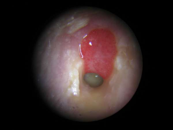
What is a cholesteatoma?
A cholesteatoma is a benign tumour that grows like an onion skin, but it is not a form of cancer. The cholesteatoma appears as a chronic purulent inflammation of the middle ear. As a subtype of chronic middle ear inflammation, cholesteatoma is also known by the outdated term otitis media epitympanalis, in which inflammatory processes occur that can result in the breakdown of bone in the middle ear. Cholestatis can lead to serious complications such as hearing loss, deafness, facial nerve paralysis or dizziness. In the worst case, damage to the meninges and/or brain is also possible. Cholesteatoma can also cause runny ears, and the secretion (foetid otorrhoea) may smell unpleasant. Cholesteatoma is diagnosed by an ENT specialist and always requires surgery. Children are more likely than average to develop cholesteatoma, although in principle it can occur at any age.
What are the causes for the development of a cholesteatoma?
In a healthy ear, the keratinising squamous epithelium, which is located in the external auditory canal, and the mucous membrane in the middle ear are completely separated from each other. However, if this natural barrier is compromised, for example by a prolonged negative pressure in the middle ear caused by a prolonged middle ear infection, it is possible for the squamous epithelium to grow into the middle ear space, spreading here and causing inflammation.
What are the different types of cholesteatoma?
Doctors distinguish between the following three types of cholesteatoma:
- Primary cholesteatoma (retraction cholesteatoma): In this form, so-called retraction pockets develop in the eardrum due to a chronic disorder of tube venitlation.
- Secondary cholesteatoma: This is the most common form of cholesteatoma and is characterised by a defect at the edge of the eardrum, which can be caused by a chronic middle ear infection in which the squamous epithelium grows into the middle ear.
- Congenital cholesteatoma: belongs to a rare type of cholesteatoma and forms due to cellular disruption during the embryonic phase. Unlike primary and secondary cholesteatoma, congenital cholesteatoma forms when the eardrum is intact.
What symptoms does a cholesteatoma cause?
At the beginning of the disease, a cholesteatoma is rather inconspicuous. Afterwards, the typical symptoms of a chronic middle ear infection can develop. These include
- Ear pain, such as ear pressure,
- Runny ears (otorrhoea), possibly with the discharge of an unpleasant-smelling fluid,
- decreased hearing, accompanied by a loss of balance,
- Dizziness, nausea and/or vomiting
If the cholesteatoma is advanced, it can cause a chronic form of otitis media, chronic bone suppuration. In this case, it is possible that the ossicles in the middle ear and the skull bones are damaged and the hearing ability is limited as a result. This can even lead to hearing loss, which can occur with eye tremor (nystagmus). In an even later stage of the disease, the facial nerve, the meninges and/or the brain can also be attacked. This can result in facial paralysis, fever, severe headaches, clouding of consciousness, a stiff neck and seizures.
How is a cholesteatoma diagnosed?
A cholesteatoma is diagnosed by the ENT doctor by means of a so-called ear microscopy as well as the examination of hearing and balance. A typical finding is a defect in the pars tensa or the pars flaccida of the eardrum, which is accompanied by whitish-yellow scales or a cell mass. Bacteriological and neurological examinations can also be performed. Computer tomography (CT) is used to precisely locate the cholesteatoma and to visualise its dimensions. Based on this knowledge, a precise treatment method can be worked out.
How is a cholesteatoma treated?
A cholesteatoma must always be removed surgically. This involves accessing the ear through an incision behind the pinna. The doctor will remove all the tissue destroyed and infiltrated by the cholesteatoma and then reconstruct the destroyed area, such as the defective ossicular chain, through a procedure called tympanoplasty, in order to restore or improve sound transmission.
The operation of a cholesteatoma is performed in hospital under general anaesthesia and lasts on average between three and four hours. After the operation, the patient stays in hospital for at least two more days. In preparation for the operation, the ENT doctor may prescribe antibiotics in the form of ear drops or tablets. These are meant to kill the bacteria and reduce the inflammation.
What is the aftercare for a cholesteatoma?
It can take up to several weeks for the wound to heal after surgery. During this time, you may experience ringing or noises in your ears. Patients should avoid getting water in their ears, especially in the first few months after the operation. Air travel, sports and saunas should also be avoided. It is possible that a tumour will reappear about six months after the operation, which can be treated with medication if necessary. In any case, it is advisable for patients to have regular check-ups and hearing tests by an ENT specialist.
Hearing usually improves completely after the successful removal of the cholesteatoma. However, it is also possible that complications occur, which manifest themselves in ringing in the ears (tinnitus) or running in the ears. Rather rarely, paralysis of the facial nerve, increased scarring or the appearance of taste disorders may also occur. In a very small number of patients, it will not be possible to reconstruct hearing, even in spite of surgery. In this case, patients will be dependent on a hearing aid.
What is the prognosis of a cholesteatoma?
A benign cholesteatoma has excellent chances of recovery. Here, as with other diseases, the earlier the cholesteatoma is diagnosed and treated, the better the prognosis.
