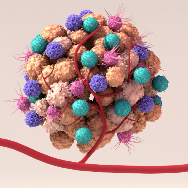
What is a chordoma?
A chordoma is a benign tumour which occurs particularly frequently in the area of the coccyx or the base of the skull (clivus). However, they can generally form anywhere along the spine and can also manifest in the bones of the mobile sections of the spine or in the sacrum, for example. Despite their benign nature, chordomas tend to have an aggressive infiltrative growth. This means that even after complete removal of the tumour, the chordoma can recur (high recurrence). Chordomas originate from the remains of the primordial spine, the so-called chorda dorsalis. Although they have grown outside the meninges, they can often lead to a compression of the brain stem and cranial nerves and accordingly cause symptoms in this area as well.
How do chordomas form?
Chordomas develop from the tissue cells of the so-called notochord. This is an embryonic structure that is involved in the formation of the spinal column. Normally, the notochord regresses in the eighth week of the foetus. However, some notochord cells may remain in the bones of the spine and the base of the skull. It is rather rare for these notochord cells to form into the cancer type chordoma. Why this happens in some people has not yet been clarified beyond doubt by medical experts.
Who gets chordoma most often?
Doctors assume that a chordoma develops due to a genetic predisposition. For example, it has been found that families with hereditary chordomas have another copy of the brachyury gene. But children who suffer from the hereditary disease tuberous sclerosis also tend to develop chordomas more often. This can be explained by the alteration of the TSC1 and TSC2 genes, which have changed due to tuberous sclerosis.
What are the different types of chordoma?
Chordomas can be divided by doctors into four subtypes, which are differentiated by their appearance under the microscope as follows:
- the conventional (classical) chordoma: This subtype occurs most frequently and consists of a type of cell that resembles notochord cells and externally resembles a chondroid dedifferentiated chordoma.
- the poorly differentiated chordoma: This subtype was only recently discovered and is distinguished from the classic chordoma by its aggressive and rapid growth. In addition, the low-differentiated chordoma lacks the gene INI-1. Children and adolescents are particularly likely to develop this subtype. However, it is also frequently found in skull base tumours.
- thededifferentiated chordoma: This subtype is even more aggressive in its growth compared to the two forms of chordoma already mentioned. In addition, the dedifferentiated chordoma tends to form metastases than conventional chordomas, for example. As with the poorly differentiated chordoma, the dedifferentiated chordoma also lacks the INI-1 gene. Almost 5 percent of all chordoma patients develop this subtype. Most of them are children.
- chondroid chordoma: This is a rather outdated term for a chordoma and dates back to the time when doctors still had difficulties differentiating between a conventional chordoma and a chondrosarcoma. Nowadays, this differentiation is no longer a problem due to the detection of the deviation of the Brachyury gene. This is because chondrosarcomas, unlike chordomas, do not have this abnormality.
What symptoms does a chordoma cause?
A chordoma usually manifests itself through compression of the brain stem and cranial nerves and can cause the following symptoms:
- Cranial nerve interference, which can cause double vision or visual disturbances in the form of reduced visual acuity or visual field loss,
- Pituitary insufficiency (hormone deficiency), which leads to general fatigue and increased need for sleep
- Headaches, nausea and/or vomiting caused by the tumour growing into the brain stem and the consequent accumulation of cerebrospinal fluid
- Destruction of the joints between the spinal column and the head area, resulting in pain with every movement of the head
How is a chordoma diagnosed?
If a chordoma is suspected, the doctor will first perform a magnetic resonance imaging (MRI), with which the chordoma can be well visualised as a hyperintense structure, especially in the T2-weighted sequence. After administering a contrast agent, the chordoma will also become visible in the T1-weighted sequence. In addition to MRI, a CT scan with a bone window can be performed to better visualise the bony destruction. This CT scan is particularly important for tumours located in the cranio-spinal junction. This is because a CT scan can be used to evaluate stability.
How is a chordoma treated?
Chordomas hardly respond to radiotherapy or chemotherapy, which is why complete surgical removal of the tumour is the goal. This is possible, at least for tumours in the midline, by endoscopic endonasal surgery. However, if the tumour is larger, it is usually difficult to remove it completely. Even if no residual tumour can be seen on the MRI after the operation, the chordoma is still present in the connective tissue nerve sheaths or in the adjacent bony structures. For this reason, it is advisable to perform proton or carbon ion irradiation after each operation. Since there is still a risk that the tumour will form again, the patient should attend regular check-ups.
What are the prognosis prospects for a chordoma?
The prognosis for a complete cure always depends on many different factors. These include the age, but also the general health of the patient as well as the type of tumour, its size and location. On average, the probability of survival with the right treatment is ten years or more.
| Pathogen | Source | Members - Area |
|---|---|---|
| Chordoma benign | EDTFL | As a NLS member you have direct access to these frequency lists |
| Chondroma | EDTFL | As an NLS member you have direct access to these frequency lists |
