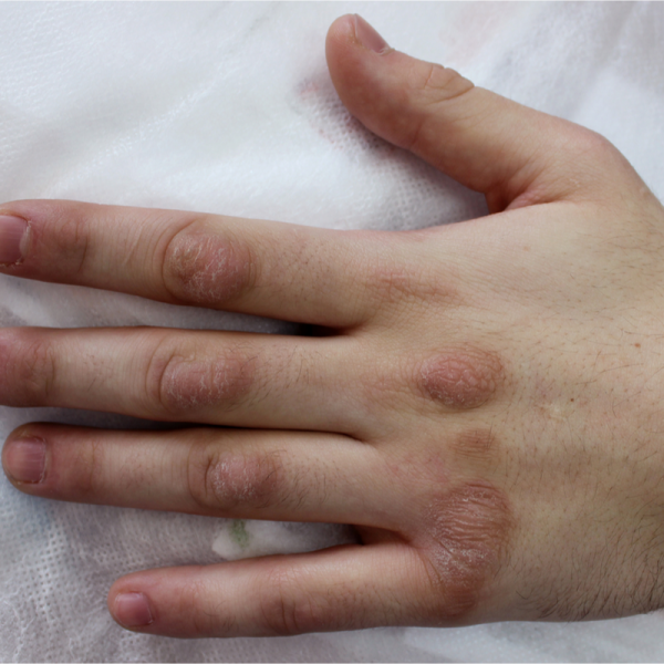
What is fibromatosis?
Fibromatosis is a skin outgrowth, or more precisely, a benign growth of connective tissue. Connective tissue includes the loose to tight tissue consisting of the intercellular mass, fatty tissue, collagen and the supportive and protective covering that surrounds the muscles and organs. Fibromatosis can be either superficial or deep and can occur in different parts of the body. It can damage neighbouring skin structures as the connective tissue tries to repair the resulting tissue damage itself, proliferating beyond the corresponding skin site.
In some patients, fibromatosis is hereditary. Although fibromatosis is benign, it can sometimes grow very aggressively and ulcer-like, invading the surrounding tissue and damaging important cell structures here. In this case, there are rarely if ever changes in the tissue cells (so-called degenerations). Fibromatosis often affects blood vessels, nerves and the lymphatic system.
How does fibromatosis develop and what are the causes of the disease?
In many cases, so-called myofibroblasts are involved in the development of fibromatosis. These are cells that move between the muscle cells (myoblasts) and the connective tissue cells (fibroblasts). They can be involved in causing proliferation in the collagen connective tissue.
Fibromatoses can be caused by inflammations or injuries, but also by external force. In many cases, however, no clear cause for fibromatosis can be identified. Some forms of fibromatosis may be congenital or inherited.
What are the different forms of fibromatosis?
Doctors distinguish between superficial and deep fibromatoses. The following clinical pictures belong to the superficial fibromatoses:
- aggressive fibromatosis, which appears on the fascia of the muscle (desmoid tumour),
- Fibromatosis colli, which often occurs in small children and can result in a torticollis,
- Fibromatosis of the gums (Jones syndrome)
- Fibromatosis occurring in the hand (Dupuytren's disease),
- plantar fibromatosis of the sole of the foot (Ledderhose's disease),
- Connective tissue disease of the penis (Peyronie's disease),
- Nodularity of the upper extremities (nodular fasciitis),
Superficial fibromatosis occurs mainly on the underside of the feet or the palms of the hands. Nodular fasciitis (fasciitis nodularis) is one of the most common superficial fibromatoses, which is mainly manifested by the formation of individual growing nodules.
The following clinical pictures belong to the deep fibromatoses:
- Fibromatosis that appears in the posterior abdomen and the lower back (Ormond's disease)
- a hardening inflammation of the connective tissue which occurs in the mediastinum of the chest (sclerosing mediastinitis)
- inflammation of the small intestine, which mainly affects the connective tissue that is permeated by fatty tissue (sclerosing mesenteritis),
What are the symptoms of fibromatosis?
The symptoms of fibromatosis depend on where exactly it is located. For example, if it is a superficial fibromatosis, such as Ledderhose's disease or Dupuytren's disease, it can often cause a feeling of pressure and traction, as well as pain and/or irritation. In some cases, smaller or larger nodules may also form. Deep fibromatoses can cause pressure pain, shortness of breath (especially in sclerosing mediastinitis) as well as back pain and kidney damage (especially in Ormond's disease).
In order to diagnose fibromatosis as early as possible, skin or tissue changes should be clarified by a doctor.
How is fibromatosis diagnosed?
Because other diseases can also be involved, it is not always easy to diagnose fibromatosis. It is typical for fibromatosis that the tissue changes cannot be clearly distinguished from the surrounding tissue. However, in order to diagnose fibromatosis beyond doubt, a biopsy must be performed. In most cases, the growth is removed during the surgical removal of the tissue sample. Within the clinical examination, it is determined whether the tissue is a benign or a malignant tumour. The degree of malignancy of the growth is also determined. If it is a malignant tumour, the doctor speaks of a fibrosarcoma and no longer of fibromatosis.
In the case of a fibrosarcoma, further examinations are necessary. In addition to a blood test, the internal organs are also examined in order to be able to plan the further treatment as best as possible. Imaging frahling procedures such as a magnetic resonance imaging (MRI) or a computer tomography (CT) can show whether the growth has already spread further or even formed metastases.
How is fibromatosis treated?
The type of treatment depends on where the fibromatosis is located, whether and what symptoms it causes, and how fast it is growing. As a rule, surgical removal of fibromatosis can be recommended.
If it is an aggressive fibromatosis, it can usually only be treated in a delaying manner. In the case of surgical removal, the healthy tissue around the fibromatosis must also be generously removed. If the complete removal of the diseased tissue is not possible, in many cases the disease will develop again after some time (recurrence). In this case, the doctor usually advises additional radiation therapy, which is carried out locally and is intended to destroy the diseased cells. However, as with an operation, the healthy tissue is also damaged by the intervention.
What complications can occur with fibromatosis?
In some patients, fibromatosis can turn out to be malignant. Specifically, this means that the diseased cells displace healthy tissue. This can lead to metastases. But it can also lead to organ infestation and, in the worst case, even organ failure. However, such courses of the disease occur rather rarely in fibromatosis.
