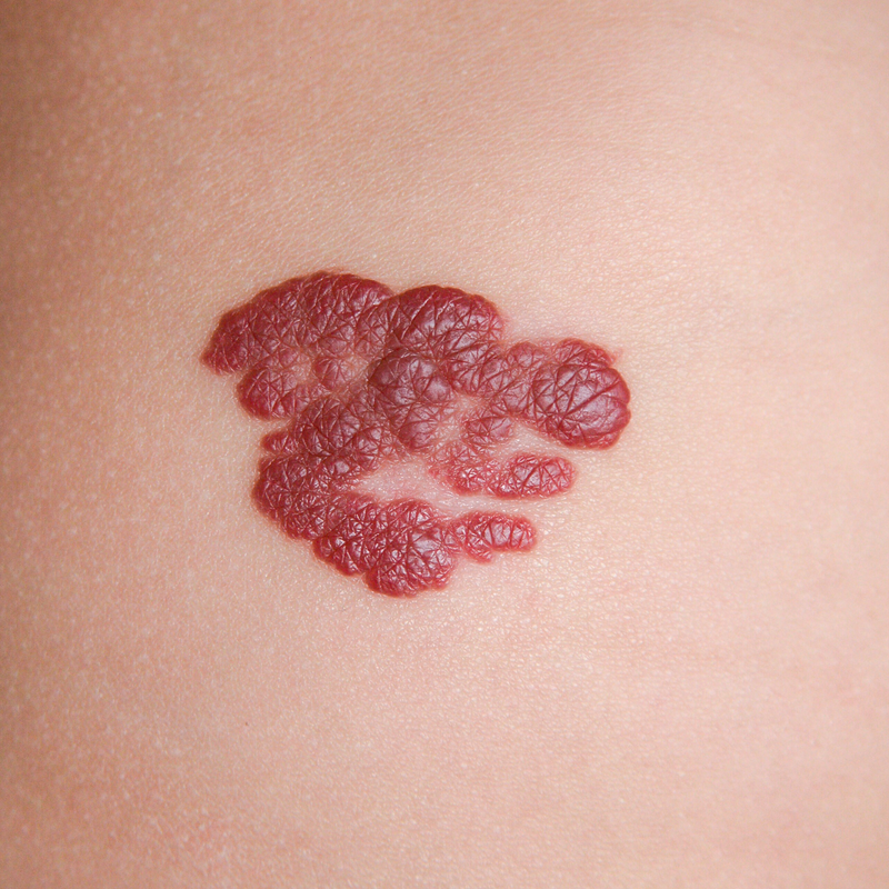
What is a haemangioma?
A haemangioma is a benign tumour of the blood vessels, which is also colloquially known as a blood sponge or strawberry stain. Haemangiomas are neoplasms, i.e. new formations of body tissue, which are caused by defective cell growth and are still very small if they are congenital. In some cases, haemangiomas can increase in size during the first years of life, then stop growing and eventually regress on their own.
Haemangiomas are among the most common types of tumours that occur in childhood and are congenital in most cases. Between 3 and 5 per cent of all babies are affected by a haemangioma, with premature babies being up to 10 times more likely to develop a haemangioma. Girls are about two to three times more likely to suffer from a haemangioma than boys. Many haemangiomas regress on their own and usually do not degenerate.
How do haemangiomas develop?
A haemangioma is caused by the formation of proliferating blood vessels, i.e. blood vessels that have the character of a tumour. The exact causes of this tumour character are still unknown to doctors. However, they assume that there are genetic factors and hormonal control.
Where do haemangiomas mainly occur?
In principle, haemangiomas can occur all over the body, but they can also affect internal organs. In particular, about one third of all haemangiomas are localised in the liver. In about 60 percent of all cases, haemangiomas often occur in the head and neck area, which is why they are also called head blood tumours. If whole areas of skin or extremities are affected by a haemangioma, doctors call it angiomatosis.
Which haemangiomas are differentiated?
Doctors distinguish between the following haemangiomas:
- Tufted angioma: is a vascular tumour that is either congenital or forms before the age of 5.
- Epithelioid cell haemangioma: is a vascular tumour of the skin.
- Glomeruloid haemangioma: is also a vascular tumour affecting the skin.
- Infantile haemangioma: is one of the most common benign vascular tumours that occur in childhood and are either congenital or appear in the first few weeks of life.
- Congenital haemangioma: vascular tumour which is in turn divided into a rapidly regressing congenital haemangioma (RICH) and a non-regressing congenital haemangioma (NICH).
- Microvenular haemangioma: occurs mainly on the extremities of young adults and is characterised by its small, reddish-brown plaques.
- Papillary haemangioma: is a vascular tumour of the skin.
- Spindle cell haemangioma (SCH): is a benign vascular tumour of the dermis, which occurs rather rarely.
- Targetoid haemosiderotic haemangioma (hobnail hemangioma): is a rather rare haemangioma which can occur at any age.
How can you recognise a haemangioma?
A haemangioma may be visible as a reddened lump on the surface of the skin. If the haemangioma penetrates into deeper layers of the skin, it may appear as a bright red or blue-red flat lump. Infantile haemangioma in particular progresses through the following three growth phases:
- 1. Growth phase: some infantile haemangiomas are not yet visible at birth and only appear after about four weeks as a white, red or blue-white spot with a dilated blood vessel visible in the centre. The infantile haemangioma can grow very quickly in the following three to four months and stop growing somewhat in the following six months, but in comparison to the growth of the body it is still disproportionately above the average of regular growth.
- 2. Stagnant phase: In this growth phase, the infantile haemangioma can remain unchanged in its growth and size for several months or even years.
- 3. Regression phase: In the regression phase, the infantile haemangioma may initially change its colour from an intense red to a grey-red or grey-skinned tone. In addition to its colour, the haemangioma also changes its shape and becomes softer and flatter. Depending on whether it is a superficial haemangioma or one that penetrates into deeper layers of the skin, the regression phase can last for different lengths of time. As a rule, almost 50 percent of all infantile haemangiomas regress spontaneously by the fifth birthday. in contrast, 70 per cent of all infantile haemangiomas have disappeared by the seventh birthday and 90 per cent by the ninth birthday.
How is a haemangioma diagnosed?
If a haemangioma appears externally on the skin, it can usually be easily diagnosed by a specialist. Nevertheless, it is important for the treatment to determine what kind of haemangioma it is. The following three aspects can be informative:
- 1. Has the haemangioma been present since birth? - If this is the case, there is a lot to be said for a vascular malformation or a congenital haemangioma.
- 2. Did the haemangioma expand in size from the 6th to the 9th month after birth? - If so, this could all point to an infantile haemangioma.
- 3. Has the haemangioma reduced in size over time? - If this is true, there is a lot to be said for an infantile haemangioma.
How is a haemangioma treated?
If it is a simple infantile haemangioma, it usually does not need to be treated at all, because it will disappear by itself over time. However, it is still advisable to have an uncomplicated infantile haemangioma checked regularly so that appropriate treatment can be initiated if necessary.
If, on the other hand, a haemangioma has appeared on the face or in the anogenital region, it is necessary to treat the haemangioma. In most cases, the haemangioma is treated with propranolol over a period of 6 to 12 months. For smaller and flat infantile haemangiomas, laser therapy using dye lasers or flash lamps can also be used. In rare cases, the haemangioma is removed surgically. This can be the case, for example, if it is located directly on the tip of the nose.
What complications can occur with a haemangioma?
Small haemangiomas, which tend to grow slowly and occur mainly in the trunk area or on the extremities, are rather uncomplicated. Fast-growing haemangiomas, which appear especially on the skin surfaces and which are in constant contact with healthy skin, such as in the armpit area, can develop complications in the form of open wounds, pain or bleeding. Particularly large haemangiomas that occur on the extremities are also suspected of affecting the growth of the affected region in children.
Above-average-sized haemangiomas that appear in the face and neck area mainly lead to aesthetic and functional limitations. A haemangioma of the eyelid, for example, can obstruct the opening of the eye and in extreme cases even lead to permanent amblyopia. A haemangioma of the mouth can lead to difficulties in eating, a permanent deformation of the lips or even an abnormality in the position of the jaw and teeth. A haemangioma on the nose can lead to either a nasal deformity or discomfort when breathing through the nose. Strongly perfused haemangiomas on the ears often lead to cartilage deformation or excessive ear growth.
