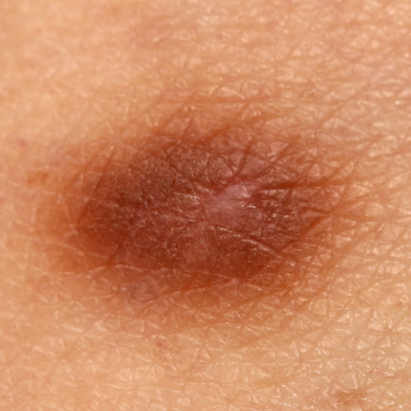
What is a histiocytoma?
A histiocytoma is a benign growth of connective tissue. Because it resembles a hard fibroma, a histiocytoma is also called a dermatofibroma. A histiocytoma usually appears as a 0.5 to 2 cm large nodule that protrudes above the skin level and takes on a brownish to red colour. A histiocytoma tends to spread deeper when the affected area of skin is squeezed. In most cases of the disease, a histiocytoma forms on the legs and especially on the lower legs. However, it can also occur on the arms. Histiocytomas are often caused by insect and/or mosquito bites. However, it can also develop after a small injury. Adults between the ages of 30 and 60 have an above-average incidence of histiocytoma. Women are affected more often than men. The disease is less common in children.
What types of histiocytoma are there?
In medicine, the term histiocytoma is used to distinguish between the following two tumours:
- Benign cutaneous fibrous histiocytoma (so-called dermatofibroma): refers to a benign skin tumour which consists of fibrocytes and collagen fibres and can develop at any stage of life, but is most common in young adults.
- Malignant fibrous histiocytoma: This includes both skin tumours and tumours of soft tissue and bone.
Why does a histiocytoma develop?
The exact cause of a histiocytoma is not yet completely clear to doctors. However, they suspect that a histiocytoma can develop as a result of an insect bite, for example from a tick. In addition, the following other possible causes of development are considered:
- as a scar after a skin injury,
- as an inflammation of a hair follicle (folliculitis),
- as a result of a minor injury
On which parts of the body do histiocytomas form?
Histiocytomas can generally occur on any part of the body, but they are particularly common on the extremities, especially the arms and legs. Histiocytomas on the palms of the hands and the soles of the feet are also described in the scientific literature, but they occur rather rarely.
How can you prevent a histiocytoma?
In general, it is not possible to prevent the development of a histiocytoma. However, you can reduce the risk of developing a histiocytoma, for example, by treating skin injuries carefully. Also, insect bites should not be scratched open and the development of hair follicle infections or skin infections should be prevented.
Are histiocytomas dangerous?
From a medical point of view, histiocytomas are not dangerous. However, they can be perceived as disturbing, especially from an aesthetic point of view. They can also interfere with movement if they are located on areas of the skin that are often exposed to friction. These include the underarm area, the lower part of the female breast or the legs.
In some cases, it is also possible for a histiocytoma to become inflamed. This is especially the case if the nipples are so-called pedunculated and twisted. They can then take on a black colour and die off due to inflammation.
What symptoms does a histiocytoma cause?
Histiocytomas appear as painless small nodules on the skin and are more common on the extremities. They can range in size from 0.3 to 1.5 cm and appear either singly or in multiples. If they contain the pigment haemosiderin, they take on a brownish to bluish discolouration. If they consist of fatty deposits, they can take on a reddish or yellowish colour.
Apart from its external appearance, a histiocytoma does not cause any other symptoms unless it impairs movement due to its location. A histiocytoma does not cause pain, nor is it prone to itching or degeneration.
How is a histiocytoma diagnosed?
A dermatologist can diagnose a histiocytoma by looking at its typical appearance. At the same time, the type of histiocytoma is determined.
How is a histiocytoma treated?
Since a histiocytoma is usually only a harmless skin change, no treatment is usually necessary. However, if the histiocytoma interferes with movement because of its location or if it is frequently inflamed, it can be removed by surgery. Surgical removal may also be advisable for the so-called pedunculated nipples, because depending on their position, they can easily become inflamed or get caught in necklaces or the like.
However, a scar remains after this procedure. Removal by CO2 laser is also a possibility. The removal of a histiocytoma is usually done under local anaesthesia and requires a few stitches to close the wound. In about five per cent of all cases, histiocytomas form again after surgical removal.
Can you remove a histiocytoma yourself?
In any case, you should refrain from removing a histiocytoma yourself. Also, a histiocytoma, for example in the case of the so-called pedunculated wart, should not be ligated on one's own or a fibroma patch applied without medical supervision. Any attempt at self-treatment carries the risk of infection, inflammation and scarring.
What home remedies can help against a histiocytoma?
Instead of surgical self-treatment, the use of home remedies can be effective in removing histiocytomas in some cases. Some sufferers drizzle apple cider vinegar on the histiocytoma several times a day. Alternatively, cotton wool soaked in apple cider vinegar can be applied to the histiocytoma several times a day. In some cases, this self-treatment has succeeded in making the histiocytoma disappear. However, it is important to make sure that the skin around the histiocytoma does not become inflamed.
