What is a myxoma?
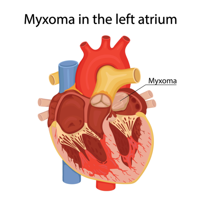
A myxoma is a rare benign cell proliferation of the heart . In about three quarters of all cases, the myxoma forms in the atrium of the heart from (so-called atrial myxoma), whereby it is located significantly more often in the left atrium than in the right atrium and sits on the inner wall of the heart. The so-called atrial myxomas form more frequently in women than in men and usually occur spontaneously in the second half of life. The peak age of onset is between the ages of 40 and 60. The myxoma arises from the atrial septum and forms from connective tissue cells. The myxoma predominantly takes on a pedunculated form and grows from the inner wall of the heart into the cavity of the atrium.
What are the different forms of myxoma?
The following forms of myxoma are distinguished:
- Atrial myxoma: This is a benign, usually pedunculated tumour of the heart. An atrial myxoma consists of an unformed connective tissue-like, mucous-like basic substance and grows from the inner wall of the heart into the cavity of the atrium.
- Ovarian myxoma: is a rare benign ovarian tumour, which consists of a mucous or gelatinous consistency.
- Odontogenic myxoma: is a rare benign odontogenic tumour.
What symptoms can a myxoma cause?
By the stalk-like connection of the myxoma of the inner wall of the heart into the cavity of the atrium, it can cause obstruction of the heart's leaflet valves . The leaflet valves connect the atrium to the ventricle. A protrusion of the myxoma can cause the following symptoms:
- Difficulty in filling the ventricle, which can lead to interruption of blood supply and possibly even to short-term collapse,
- Heart palpitations,
- Shortness of breath,
- Irritable cough,
- Chest pain,
- Anaemia,
- unwanted weight loss,
- persistent fatigue,
- Blood count changes with increased inflammation values.
The
complaints can develop depending on the position.
For example, some patients may experience
greater problems when lying down than when standing up. General tumour symptoms such as anaemia,
weight loss and fatigue can also occur, as can a
change in the blood count and increased inflammation levels.
Complications can also occur despite the benign nature of the myxoma, which can be explained
in particular by the location of the tumour. In addition to
cardiac arrhythmias, the possible development of
blood clots in the atrium should be mentioned here. In the worst case, these can lead
to pulmonary embolisms, but also to strokes, which
are potentially life-threatening and can even be fatal
.
What causes a myxoma to form?
Myxomas, which mainly develop at a younger age, can be caused by an inherited symptom complex (Carney complex). The Carney complex causes tumours to form on the heart, breast and skin as well as specific moles. Hormonal dysfunctions of the adrenal cortex can also occur due to the Carney complex.
A myxoma can also develop due to a clinical syndrome, such as a Mazabraud syndrome or a Carney complex .
What complications can arise from a myxoma?
In about 50 percent of all cases, cardiac arrhythmias occur due to a myxoma . But blood clots, a so-called thrombus, which can form around the myxoma, are also potentially dangerous. This is because in about 25 percent of all cases, a blood clot can lead to an embolism if it is flushed out with the blood and, in the worst case, even cause circulatory problems of the brain or sudden brain death. Vascular occlusion in the legs or pulmonary oedema can also occur as complications of a myxoma.
How is myxoma diagnosed?
The diagnosis of a myxoma can be difficult because of the mostly unspecific symptoms, especially since many myxomas do not cause any symptoms for a long time. It is therefore possible that a myxoma is initially misdiagnosed or discovered by chance during an ultrasound examination of the heart. The following examinations should therefore be carried out in order to make a reliable diagnosis :
- physical examination: If there are unusual heart murmurs, these can indicate a disturbed blood flow, which can occur due to a faulty valve closure of the heart.
- echographic imaging of the heart: should normally be able to indicate a myxoma, as changes within the atria can be made visible through the examination .
- other imaging procedures such as a magnetic resonance imaging (MRI) of the heart and/or a computed tomography (CT) scan can usually make a myxoma visible.
How is a myxoma treated?
If possible, a myxoma should be surgically removed because of its sensitive location and the serious complications it can cause. Before the surgical procedure, the patient is often prescribed an anticoagulant to prevent the formation of a blood clot. In any case, an attempt should be made to surgically remove the entire tumour. During the operation, the patient is connected to a heart-lung machine, which acts as a bypass circuit and continues to oxygenate the blood by machine despite the heart being stopped .
The postoperative recovery process can take some time, depending on the patient. After the operation, it is highly recommended to attend the regular check-ups to check whether new myxoma tissue has formed.
What is the prognosis for myxoma?
The prognosis always depends on how deeply the myxoma has already grown into the surrounding tissue and whether it was actually possible to remove it completely through the operation . If the tumour could be completely removed, the prognosis is very good. However, the chances of recovery depend on whether several myxomas have occurred and whether a functional disease of the heart has already occurred due to the tumour.
