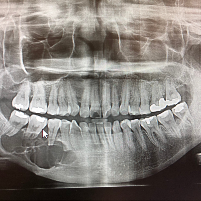
What are odontogenic tumours?
Odontogenic tumours are rare neoplasms of the articular cartilage and bone. They form predominantly from embryonic remnants of the tissue involved in tooth formation (odontogenic) and form exclusively in the jaw bone or the freely movable oral mucosa (alveolar mucosa). Most odontogenic tumours are benign (bengine) neoplasms. . Occasionally, however, malignant tumours such as sarcomas or carcinomas can also develop, which are odontogenic in origin. In 90 percent of all cases, odontogenic tumours develop between the 6th and 20th year of life.
What forms of odontogenic tumours are distinguished?
Odontogenic tumours can be classified as follows:
- hamartomatous tumours: this includes tumour-like, benign tissue changes. They form due to dispersed or defective germ tissue .
- non-neoplastic changes: this includes abnormal cell changes.
- metastatic malignant neoplasms: this includes malignant neoplasms that form metastases.
What types of benign neoplasms of odontogenic tumours are there?
Benign neoplasms of odontogenic tumours can be well defined macroscopically and show a locally displaced growth. The benign tumour forms can usually be removed without damaging the surrounding region . There is usually no need to worry about any functional restrictions either . Doctors distinguish between so-called extirpation, i.e. the complete surgical removal of the tumour and excision, i.e. cutting the tumour out of the affected tissue.
- Ameloblastoma:The classic form of an ameloblastoma occurs within the bone (intraosseous), spreads to the neighbouring structures and has a destructive effect on them . The more rarely occurring ameloblastoma variants include Unicystic ameloblastoma, Peripheral ameloblastoma (also called extraosseous ameloblastoma of the soft tissues) and Desmoplastic ameloblastoma. Approximately 20 percent of all odontogenic tumours are ameloblastomas, which usually occur between 30 and 50 years of age.
- Ameloblastic fibroma: This is a rare, benign form of tumour that often appears at with an unerupted tooth.
- Adenomatoid odontogenic tumour (AOT) (also called adenoameloblastoma): is also a benign form of tumour that occurs due to defectively differentiated or scattered germ tissue (hamartomatous). The adenomatoid odontogenic tumour occurs in 58 to 63 percent of all cases in the African and Asian region.
- Fibromyxoma (also called odontogenic myxoma):occurs relatively rarely.
- Calcifying epithelial odontogenic tumour (KEOT) (also called Pindborg tumour):is also a rare form of tumour.
- Calcifyingodontogenic cyst (also called Gorlin cyst):occurs relatively rarely, i.e. in only 2 percent of all odontogenic tumours, and forms as a cyst.
- Odontoma: usually forms near a retained tooth. Doctors distinguish between two types: a complex odontoma and a compound odontoma. While a complex odontoma contains all tooth-forming tissues mixed together, a compound odontoma consists of the smallest rudimentary tooth structures. Odontomas are among the most common odontogenic tumours and account for approximately 73 per cent of all hamartomas in North America and Europe.
- Odontogenic fibroma:occurs rarely and can occur in different morphological variants.
- benign cementoblastoma (also called true cementoma):also occurs rarely and forms from the cementum-forming cells of the tooth.
What types of malignant neoplasms of odontogenic tumours are there?
Malignant neoplasms of odontogenic tumours infiltrate neighbouring structures and grow in a locally destructive manner. They also tend to metastasise and often recur (recurrence).
- Odontogenic carcinoma: is very rare and difficult to differentiate.
- Odontogenicsarcoma: is extremely rare.
What symptoms does an odontogenic tumour cause?
Odontogenic tumours usually behave asymptomatically, as they also tend to grow slowly . However, if it is a tumour form with a pronounced growth, swelling, a change in teeth, tooth root resoptions as well as tooth loosening and an increasing pressure on the mandibular nerve and associated sensitivity disorders can be the result.
How is an odontogenic tumour diagnosed?
Since odontogenic tumours usually do not cause any symptoms, they are often diagnosed as an incidental finding, for example, during a check-up, . An odontogenic tumour can be visualised using the usual imaging procedures. In order to make a definite diagnosis, a histological examination of the tumour tissue is required.
How is an odontogenic tumour treated?
As a rule, an attempt is made to surgically remove the odontogenic tumour. In some cases, it is then necessary to reconstruct the bone in the affected area. Since an odontogenic tumour tends to form again (recurrence), the tumour should be removed over as large an area as possible . In most cases, it is unavoidable to remove the affected part of the jaw (resection)
For the reconstruction of the jaw bone, a temporary reconstruction is usually made first, for example using a bridging plate (titanium plate). Later, this titanium plate is removed and the defective jaw bone is reconstructed with an autologous bone graft. For the production of the autologous bone graft, microsurgically reanastomosed bone grafts can be used. These can come from the iliac crest or the fibula, for example .
What is the prognosis for an odontogenic tumour?
The prognosis of an odontogenic tumour always depends on the stage of the disease at the time of diagnosis, but also on whether it is a benign or a malignant tumour. Furthermore, the type of odontogenic tumour is decisive. Generally odontogenic tumours tend to form again after a while (recurrences). In order to diagnose a possible tumour at an early stage and to treat it , the patient should make use of regular check-ups even after the successful treatment .
