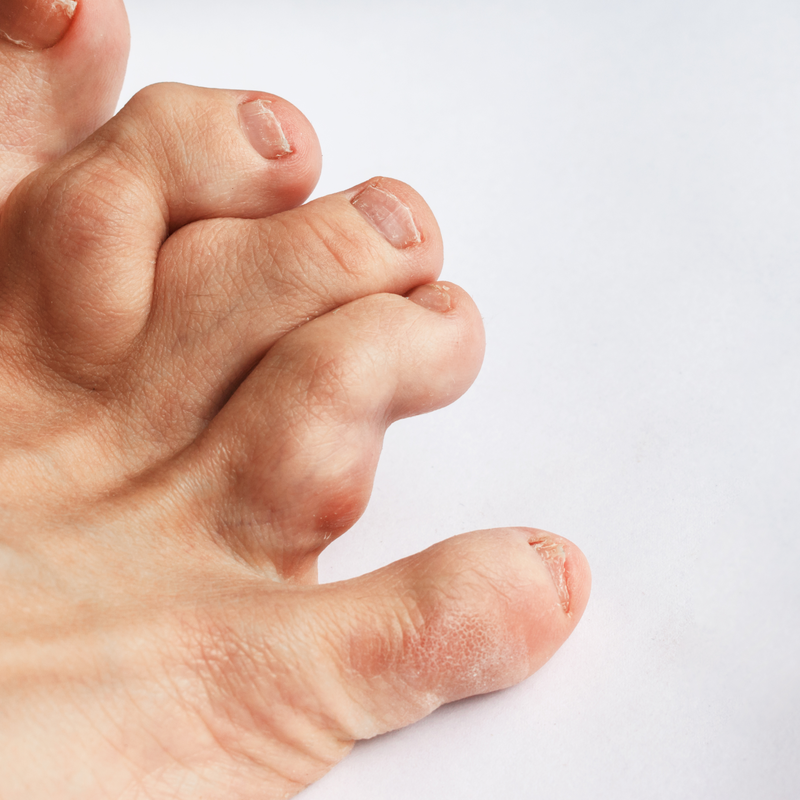
What is Ollier's disease?
Ollier's disease is a rather rare skeletal disease, also known as Multiple Enchondromatosis. It is a benign, cartilaginous tumour which can form in the medullary cavity of long tubular bones. In most cases, Ollier's disease forms near the growth plates of the bones . The bones that are most often affected are the femur, but also the humerus and the fingers and feet. Ollier's disease affects children and adolescents with above-average frequency . The tumours typical of disease, the so-called enchondromas, stop growing as soon as the bones have also completed their longitudinal growth. In most cases, Ollier's disease remains benign, but can degenerate and become malignant in almost 25 per cent of all cases .
Ollier's disease was first diagnosed in 1900 by the French surgeon Louis Leopold Ollier. The name of the disease therefore goes back to him. Ollier is considered the founder of modern orthopaedic surgery and diagnosed Ollier's disease on a 6-year-old girl who had deformities and swellings on her forearm, thigh and fingers. Nowadays, Ollier's disease is considered to be a set of pathologies known as enchondromatosis at .
How does Ollier's disease develop?
The disease arises from the formation of several benign tumours, which are also called enchondromas. These enchondromas develop mainly in the middle part of the long bones (diaphyses) and in the long bones, which are located between the diaphysis and the epiphysis (metaphyses). The formations of multiple enchondromatosis are distributed asymmetrically. However, one half of the body is always preferred. The most frequent outbreak sites affect the phalanges, the metacarpals and metatarsals as well as the humerus and/or the femur . In rather rare cases, Ollier's disease can also affect flat bones, for example the pelvis.
An enchondroma is a benign growth that develops from differentiated hyaline cartilage . In most cases, this is the remnants of embryonic cartilage that is present in the bones formed by enchondral ossification. The enchondral ossification proceeds according to the following defined stages:
- undifferentiated stem cells of the connective tissue (mesenchymal cells) form into hypertrophic chondrocytes,
- the cartilage matrix is replaced by mineralised bone.
Since
Ollier's disease forms uneven lesions, the
conclusion is that the disease arises from a postzygotic somatic
mutation, which results in mosaicism. However, medical researchers have not yet been able to find
any specific chromosomes or mutations
that can be held responsible for this phenomenon.
What causes Ollier's disease?
Doctors do not yet know what causes Ollier's disease. They also largely rule out a genetic cause. They also largely rule out a genetic mutation for the formation of the bone tumours.
What are the symptoms of Ollier's disease?
The disease only causes pain in very few cases. However, the changes caused by Ollier's disease can affect bone growth. As a result, deformities, but also the fracture of a bone in the affected area can occur.
Since Ollier's disease can degenerate in almost 25 percent of all cases, it is important to see a doctor immediately if the disease is suspected. The following signs in a child may indicate the disease:
- Growth disturbances and/or deformities,
- A tendency to frequent bone fractures
Depending
on where Ollier's disease develops, this can lead to
different complaints. For example, if the lower
extremities are affected, lameness can occur due to the shortening of the extremities
. If the tumour is at the level of the trunk, there may be
instability of the pelvis. For example, if the
base of the skull and especially the sphenoid bone is affected, the patient may complain of
headache, hoarseness, but also facial dysaesthesia and/or
hearing loss.
How is Ollier's disease diagnosed?
Since the disease is often asymptomatic, the diagnosis is often made as an incidental finding by an X-ray, computer (CT) or magnetic resonance imaging (MRI). Imaging shows enchondromas as a homogeneous mass in an elongated or oval shape and a well-defined bone margin. Existing lesions can often be located along the long axis of the bone and contain calcifications, which are visible as granular opacities.
How is Ollier's disease treated?
Ollier's disease cannot be treated with regard to its causes . In many cases, treatment is not even necessary, provided there are no deformities or bone fractures. Normally Ollier's disease is monitored by a doctor in order to diagnose possible tumour degeneration at an early stage. For the treatment of deformities or bone fractures, an operation may be necessary . This is usually aimed at removing the tumour. To do this, the bone cavity is emptied and filled with bone graft. If so-called cartilage islands remain , the disease can relapse. Surgery is not only used to remove tumours, but can also treat deformities, for example. The operation is often followed by physiotherapy. Analgesics can be used to treat pain. These are painkilling medicines.
What complications can Ollier's disease cause?
If enchondromas are present in the phalanges, they can cause severe finger deformities. However, enchondromas can also develop into malignant tumours, such as the so-called chondrosarcomas, which are particularly common in young adults. These transformations into chondrosarcomas occur more frequently than average in lesions that primarily affect flat and long bones. The development of chondrosarcomas is less frequent in small bones of the hands and feet.
